The following article comes from the international ophthalmology news, the author of the international ophthalmology news
Editor's note:Innovation is the first driving force leading development and a major proposition of today's era. The accelerated evolution of a new round of scientific and technological revolution and industrial reform highlights the urgency of accelerating the improvement of China's scientific and technological innovation ability.This is also true in the field of medical ophthalmology. The innovative research and discovery of ophthalmic diseases represent a significant improvement in the comprehensive strength of China's ophthalmology. In some fields, it even moves towards the "leading" position, which not only highlights China's hard strength of Ophthalmology, but also improves the authoritative voice of China's ophthalmology in the international ophthalmology field.At the recent aao2021 meeting, Professor Wei Wenbin's team of Beijing Tongren Hospital reported the scientific research results on the real vascular morphology of choroidal osteoma. This is the first time in the world to define the vascular morphology of choroidal osteoma. It is an extremely leading breakthrough in the international field and has attracted extensive attention of international ophthalmologists.
Choroidal osteoma, an intractable mystery in ophthalmology
In the field of Ophthalmology, choroidal osteoma is a rare disease, which mainly occurs in young and middle-aged people, mostly women, and some patients have both eyes at the same time.Although it is a benign tumor with a low incidence rate, the lesion is often located in the posterior pole of the eyeball due to its special location.At the same time, the growth of neovascularization (CNV) is often combined in the process of tumor progression. The occurrence of CNV in macular region will lead to further decline of visual function and even blindness in severe cases.At present, it is found that choroidal osteoma has some unique morphological manifestations through imaging techniques such as ultrasound, fluoroscopy and CT.However, because its essence is bone tissue, it has shielding effect on many imaging examinations. At present, it is difficult to capture its histopathological morphology in vivo.Therefore, due to the characteristics of the disease and the limitations of medical technology and examination equipment, ophthalmologists have very limited cognition on the specific morphology, internal tissue structure, characteristics between blood vessels and tissues in living state, pathogenesis and progress law of choroidal osteoma, which also brings many difficulties to its diagnosis and treatment.So far, there is no effective intervention strategy to prevent the progression of choroidal osteoma.CNV can only be found in time through OCT, and the visual function of some patients can be retained through anti VEGF treatment.
"Helpless, the feeling of watching the patient's disease progress until he becomes blind, but there is nothing to do is very painful."
It is the fearless spirit of Beijing Tongren Hospital and the firm belief inherited from Chinese ophthalmology to dare to bite "hard bones" and attack "big problems".Therefore, Professor Wei's team has always been committed to the research of choroidal osteoma, hoping to deeply understand its histological and morphological characteristics in vivo through the observation of its structure, and then explore its pathogenesis and disease progression characteristics, so as to find effective intervention methods to prevent patients from blindness.
Thousands of chisels, thousands of blows, and finally something: Choroidal osteomas are defined as sea fan vascular networks (sfvns) for the first time in the world!
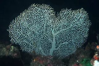
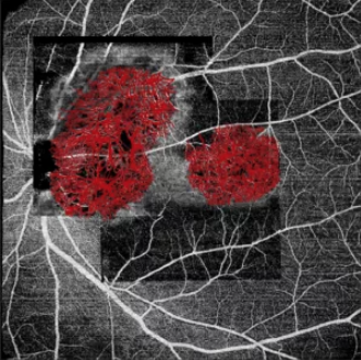
Because the bone tissue of choroidal osteoma is very dense, the refraction and shielding effects in the imaging process cause great interference to its vascular observation, which makes it difficult to clearly observe its vascular morphology by previous imaging techniques.With the breakthrough of imaging technology, with the help of new OCT equipment independently developed in China, Professor Wei and his team Dr. Zhou Nan repeatedly observed the vascular morphology of choroidal osteoma.The new OCT has strong penetration to choroidal osteoma and high resolution of vascular morphology. With the help of artificial intelligence algorithm, it finally restores the real vascular morphology of choroidal osteoma in vivo. Whether it is calcified area or decalcified area, it can clearly show the vascular morphology in the tumor.This result excited the team. Like the fog cleared, the boats sailing in the sea were clearly reflected in the eyes. It also made everyone realize that choroidal osteoma is not an irregular vascular shape as suggested by previous imaging.Choroidal osteoma is very similar to the shape of human five fingers. More detailed, its vascular shape is like a sea fan.When exploring the undersea world, I believe everyone has observed the shape of the sea fan. Although its branches are numerous and redundant, its overall shape grows regularly. Based on the root, it expands in a fan shape and finally forms a fan.The vascular morphology of choroidal osteoma is no different from that of sea fan, so Professor Wei's team vividly named it sea fan vascular networks (sfvns).This is the first time in the world to define the vascular morphology of choroidal osteoma, and this morphology is not an individual case. Professor Wei's team observed sfvns in more than 30 cases and different parts of each patient, captured the essence of choroidal osteoma, and took the lead in deepening the understanding of this disease.

scallop

The scalloped vascular network of choroid osteoma
At present, there are many mysteries about choroidal osteoma, such as:
·What causes the occurrence and growth of this tumor?
·What are the characteristics and progression of choroidal osteoma during its growth?
·What is the relationship between the occurrence of complications and the pathological changes of the tumor itself?
”Sfvns provides a powerful focus for solving these mysteries. As an important observation index in the process of disease monitoring and follow-up, sfvns helps ophthalmologists find ways to intervene the disease and prevent the serious impact of disease progression and complications on visual function."Professor Wei pointed out that sfvns provides a very good direction for further study of choroidal osteoma in the future.
Perception: the combination of medicine and industry has achieved the realm of medical science and technology innovation
Scientific research and innovation constantly promote and promote people's understanding of diseases, so as to seek better diagnosis and treatment strategies, so that many difficult eye diseases can be cured. Ophthalmic clinicians should not only undertake clinical diagnosis and treatment, but also shoulder the important task of medical scientific research and innovation. Professor Wei's team's scientific research breakthrough is a great morale and excitement for Chinese ophthalmologistsThe popular news represents that China's ophthalmology research level leads the international forefront in another new field. Referring to this breakthrough, Professor Wei said that this success reflects "medicine" and "Engineering"In clinical work, ophthalmologists summarize the clinical difficulties and bottlenecks through a large number of contacts and observation of cases, but how to deeply study its pathological characteristics, pathogenesis and intervention methods needs "work"Fang Lai provides advanced equipment and drugs to promote the cognition of diseases and seek effective solutions. The combination of medicine and work can promote the clinical development of Ophthalmology, urge clinicians to do more in-depth research, and make more difficult eye diseases "come to light". At the same time, clinical difficult problems are important to "work"The higher requirements put forward by Fang also promote the continuous upgrading, optimization and improvement of examination and diagnosis equipment to a higher level, further promote the rapid development of medical technology and bring better diagnosis and treatment services to patients. Here, I would like to thank Dr. Zhou Nan and Dr. Peng Xianzhao for their great efforts and important contributions to the breakthrough scientific research of choroidal osteoma。
The combination of doctors and workers is an important way for both sides to benefit each other, promote each other's growth and achieve each other. We look forward to bringing more breakthroughs for choroidal osteoma and other difficult eye diseases through closer and more effective combination of doctors and workers in the future, and finally benefit thousands of patients trapped in eye diseases, so that everyone can live a broad life in the bright vision!