The cornea is a transparent membrane located on the anterior wall of the eyeball. Corneal endothelial cells are located on the innermost side of the cornea, which plays a role in maintaining corneal transparency. More importantly, the number of corneal endothelial cells directly affects the occurrence of rejection reaction in the process of corneal transplantation.
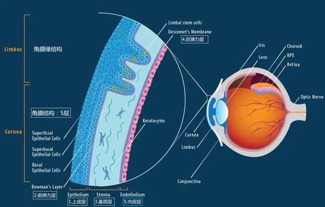
Corneal endothelial cells are very sensitive to the changes of the surrounding environment. Once they are damaged by trauma, disease or surgery, their barrier function and active operation function will be destroyed. Moreover, corneal endothelial cells are non renewable. If the loss is too large, it is easy to cause corneal endothelial decompensation, resulting in serious adverse reactions such as corneal edema and bullous keratopathy.
In recent years, due to the wide application of artificial corneal transplantation technology, corneal endothelium is often damaged, a large number of corneal endothelial cells rupture and die.
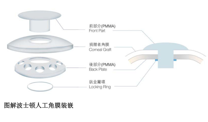
For example, the Boston artificial cornea adopts a non threaded design and does not need to rotate the front disc or rear disc to avoid scratching the donor corneal endothelial cells. However, the off-stage assembly operation of the front disc cornea rear disc combination will inevitably cause damage to the donor corneal endothelial cells.
Next, I will combine the product structure and surgical methods of Boston artificial cornea to show you the wasted life of corneal endothelial cells.
I
Boston artificial corneal transplantation is mainly divided into two steps: artificial corneal complex assembly and artificial corneal implantation.
During the assembly of artificial corneal complex, two actions of doctors will damage the endothelium of donor corneal grafts, resulting in the rupture and death of a large number of corneal endothelial cells.
First, the donor corneal graft will undergo "knife operation" twice. For the first time, the intact donor corneal graft (about 11mm) is drilled by a ring drill with a diameter of 8.5mm; The second time, the center of the 8.5 mm corneal graft was drilled by a ring drill with a diameter of 3 mm.
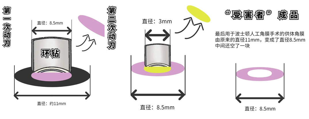
Note that continuous drilling of corneal grafts will kill a large number of corneal endothelial cells, lead to rejection, and put high-risk patients into a more dangerous situation. Moreover, with the increase of age, corneal endothelial cells will degenerate and the cell density will gradually decrease. The rupture and death of such a large area of corneal endothelial cells is equivalent to making things worse.
In addition, the action of "successively inserting the lens column, back plate and C-type titanium ring" of Boston artificial cornea will also cause corneal endothelial damage, causing corneal endothelial cell rupture and death.
This "one ring one ring" operation also has another slot point, that is, it can not completely and closely inlay the artificial cornea with the donor cornea, which is easy to cause aqueous humor leakage and endanger the normal metabolism of the lens and cornea, resulting in various lesions.
II
After the artificial corneal complex is assembled, the Boston artificial cornea can be implanted. In this process, the product structure of Boston artificial cornea once again damaged the endothelium of donor corneal graft.
During the operation, after drilling the patient's cornea with a ring drill, the doctor will fix the assembled artificial corneal complex on the implant bed with 16 stitches.
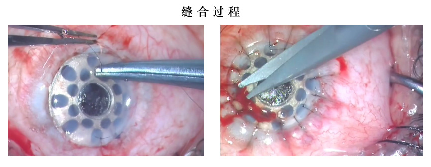
In the process of suturing, it is bound to damage the endothelium of donor corneal grafts. We should know that corneal endothelial cells are extremely sensitive to the changes of the surrounding environment. 16 silk threads enter the ocular tissue, which is easy to cause corneal endothelial injury and a large number of endothelial cell death.
At the same time, the silk thread spread all over the edge of the removed cornea will also hinder the expansion and migration of surrounding cells.
After the rupture and death of corneal endothelial cells, in order to repair the defect area, the expansion and migration of surrounding cells are needed to cover the posterior surface of cornea and form a healthy gap junction complex. Once blocked, the corneal endothelial function will be decompensated. In the case of decompensated corneal endothelial function, the hexagonal mosaic image of endothelial cells will change, which can cause bullous keratopathy in severe cases.
After the operation, the doctor will routinely place a corneal bandage lens above the artificial corneal lens column. Although this action will not cause damage to the endothelium of donor corneal graft, it is not conducive to the healing of damaged tissue in the eye.
Why do you say that? Let's look at this picture again:
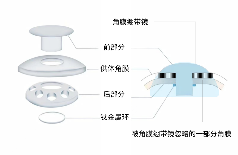
The new blue part is the corneal bandage lens, and the corneal wound in contact with it can heal faster. However, due to the umbrella shape of Boston artificial cornea lens column, the donor cornea in the black shadow cannot fit with the corneal bandage lens, which affects the healing of corneal tissue, and it happens that this part of corneal tissue needs healing most.
At present, keratopathy ranks second among blinding eye diseases, which seriously affects the quality of life of patients. The main physiological basis of blindness caused by corneal diseases is that the corneal endothelium is very "fragile".
If you think about it, the Boston artificial cornea will "ravage" such a fragile corneal endothelium three times or even many times. For the corneal endothelium, isn't it a waste of life?