Corneal epithelial lesion refers to the pathological state that the function and integrity of corneal epithelial barrier are destroyed due to various factors under the premise of normal source of limbal stem cells, resulting in partial or full-thickness loss of corneal epithelial cell layer. Clinically, corneal epithelial lesions are very common, which can be caused by a variety of causes. Corneal epithelial injury caused by different factors has different clinical characteristics. International ophthalmology news specially invited Professor Gong LAN from the eye, ear and Throat Hospital Affiliated to Fudan University to explain in detail the clinical characteristics of corneal epithelial injury caused by different factors and share the treatment strategies of corneal epithelial lesions.
Analysis: risk factors and clinical characteristics of different corneal epithelial lesions
Corneal epithelial lesions can be divided into primary corneal epithelial lesions and secondary corneal epithelial lesions.
Primary corneal epithelial lesions are rare and are mostly genetically related, such as:
1. Corneal epithelial basement membrane dystrophy (also known as map dot fingerprint dystrophy), which is due to the abnormal development of corneal epithelial basement membrane, resulting in repeated epithelial loss;
2. Corneal lattice dystrophy is characterized by irregular branching white thin strips and turbid spots in the superficial parenchyma and Bowman's layers, which gradually expand, thicken and increase, and interweave into a grid; It may also be accompanied by recurrent epithelial defects.
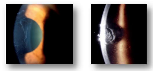
Figure 1. Primary corneal epithelial lesions. The left picture shows corneal epithelial basement membrane dystrophy, and the right picture shows corneal lattice dystrophy
Secondary corneal epithelial lesions are more common in clinic, which are common in various mechanical injuries, infections, immune factors and so on. The clinical characteristics of corneal epithelial lesions caused by different factors have their own characteristics.
1. Repeated corneal epithelial erosion, often with a history of nail, paper, plant and other abrasions. Mechanical trauma can lead to the abnormality of basement membrane junction complex, which makes the adhesion of basal epithelial cell layer become loose, and finally leads to repeated corneal epithelial exfoliation. Mechanical injury of corneal epithelium often manifests as sudden eye pain, photophobia, tears and other symptoms in the morning.
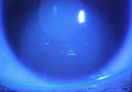
Figure 2. Structural abnormality of acquired epithelial basement membrane caused by mechanical injury, manifested as "finger cap" repeated epithelial exfoliation
2. Corneal pathogen infection can also lead to epithelial lesions. The most common is corneal epithelial lesions caused by herpes simplex virus. The virus activates and replicates in epithelial cells, causing epithelial cell necrosis and disintegration, and then epithelial abscission. The typical herpes simplex virus corneal ulcer is dendritic, terminal bifurcation and nodular enlargement.
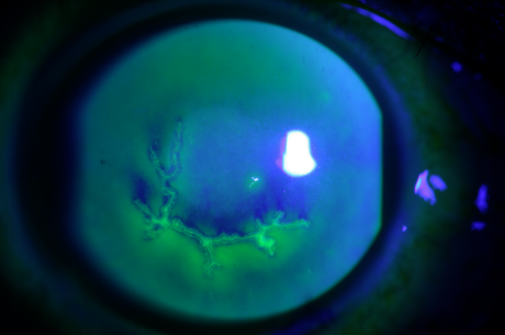
Figure 3. Dendritic ulcer of corneal epithelium caused by herpes simplex virus
3. Corneal epithelial lesion after corneal refractive surgery belongs to secondary corneal epithelial injury caused by surgery. The possible causes of corneal epithelial lesions after refractive surgery include intraoperative nerve disconnection, decreased perception and decreased expression of neurotrophic factors. The clinical symptoms of surgery induced corneal epithelial injury include: eye tingling, dryness, and abnormal corneal secretions.
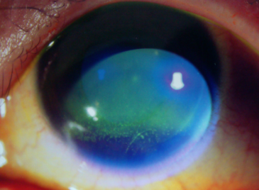
Figure 4. Case 1 year after LASIK: dense corneal epithelial exfoliation is still visible
4. Drug toxic corneal epithelial lesions often occur on the basis of primary corneal diseases, which are mostly caused by the toxic accumulation of drugs and preservatives. The typical manifestation is pseudodendritic exfoliation of epithelium, accompanied by a long-term history of eye drops.

Figure 5. Drug induced corneal epithelial damage, presenting pseudodendritic epithelial defects
5. Neurogenic corneal epithelial lesions are often caused by the damage of the trigeminal nerve, such as surgery, trauma, tumor, etc. because the sensitivity of the cornea that loses trigeminal innervation decreases, and the protective mechanism of the cornea weakens, it is easy to be damaged. At the same time, the loss of innervation leads to corneal epithelial dystrophy, and the repair mechanism is weakened. The typical clinical feature is that the signs are heavier than the symptoms, and most patients have no serious eye pain, tears and other irritating symptoms, which is related to the decline of corneal perception after trigeminal nerve injury.
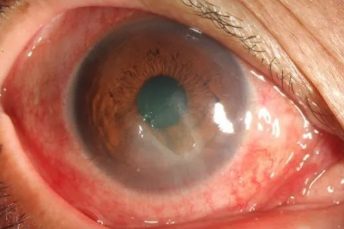
Figure 6. A 56 year old female patient complained of right eye redness, but no eye pain and tears. She had a history of trigeminal neuralgia surgery 3 years ago and was diagnosed as neurotrophic keratitis
6. Corneal epithelial lesions can also be secondary to systemic immune system diseases. Autoimmune diseases such as Sjogren's syndrome and secondary Sjogren's syndrome caused by rheumatoid arthritis are easy to cause long-term immune inflammation on the ocular surface, leading to corneal epithelial erosion.
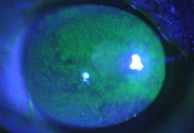
Figure 7. A 52 year old female patient has red eyes, sand sensitivity and photophobia for more than 3 years, and has a history of rheumatoid arthritis for 10 years; Schirmer test of both eyes: 0mm/5min, tear film rupture time of both eyes: 1s. It is corneal epithelial injury caused by autoimmune diseases.
7. Exposed corneal epithelial injury refers to the exposure, drying and repeated exfoliation of corneal epithelium caused by incomplete eyelid closure. Eyelid insufficiency is mostly caused by exophthalmos, ectropion, abnormal eyelid structure and facial nerve paralysis.
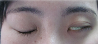
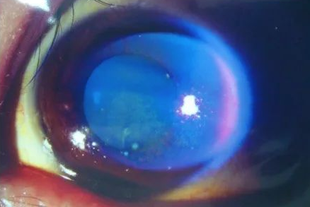
Figure 8. One year ago, the patient underwent resection of intracranial tumor due to brain tumor. After operation, he developed left facial paralysis. Recently, his left eye was dry and foreign body felt for half a month. He was diagnosed as exposed keratitis
Tracing the source: treatment strategies for corneal epithelial lesions
Professor Gong shared four strategies for treating corneal epithelial lesions:
1. Removal of etiology: corneal epithelial injury caused by different factors has different etiologies, and removal of etiology is the most important for corneal epithelial repair. For example, pathogen infection should be treated with sensitive drugs on the basis of clear diagnosis of the nature of the pathogen. Corneal epithelial damage caused by drug toxicity should be stopped or reduced in time. Neurotrophic keratitis can try to use nerve growth factor drugs, and for corneal epithelial damage caused by autoimmune diseases, attention should be paid to the systemic control of the primary disease.
2. Improve the microenvironment of epithelial repair: "ocular microenvironment" depends on tears, cells, nerves, immunity and other comprehensive factors to maintain a balance. Once one or more factors are out of balance, a series of chain reactions may occur on the ocular surface, resulting in the imbalance of ocular surface function; Most corneal epithelial injuries are accompanied by the imbalance of ocular surface microenvironment. Improving the ocular surface environment can be divided into anti-inflammatory and improving tear film. When there is a definite inflammatory reaction on the ocular surface, anti-inflammatory drugs such as glucocorticoids and immunosuppressants can be given locally; At the same time, it can give high-quality artificial tears to improve the stability of tear film and contribute to the repair of corneal epithelium.
3. Promote epithelial repair through external forces: for the treatment plan to promote corneal epithelial repair, the principle of step by step is adopted, which can be divided into "four steps". The first step is basic medication, which can provide energy, nutrition and growth factors for epithelial repair; The second step is to wear bandage glasses to provide mechanical support and protection, close the corneal wound and relieve pain; The third step is amniotic membrane covering, which is equivalent to "biological corneal contact lens", to protect the corneal epithelial defect area and create a repair space; The fourth step is eyelid fissure closure, which protects the cornea by bonding the upper and lower eyelids and avoiding exposure factors. It is suitable for other poorly treated eyelid insufficiency or neurotrophic keratitis.
4. Prevention of infection: corneal epithelial damage weakens the ability of the cornea to resist pathogens. Extensive epithelial erosion may secondary to corneal infection, causing serious consequences. At the same time of repairing corneal epithelium, giving local antibiotic eye drops to prevent corneal infection is an important adjuvant treatment.
summary
Corneal epithelial injury is a common ophthalmic disease. Its etiology can be divided into mechanical injury, pathogen infection, post refractive surgery, drug toxicity, neurotrophic, immune and exposure. The diagnosis of corneal epithelial injury depends on the history and typical symptoms and signs. Its treatment strategies mainly include removing the cause, reconstructing the ocular surface microenvironment, promoting corneal epithelial repair and preventing infection. Correct diagnosis and intervention of corneal epithelial injury types are helpful to repair corneal epithelium, reduce patients' symptoms and improve prognosis.