Research team: Peking University Third Hospital Eye Center, Wuhan Institute of Virology
Foreword: In recent years, with the widespread application of molecular biology techniques in ophthalmology clinics, ophthalmologists have discovered that cytomegalovirus (CMV) is closely related to eye diseases, and Asian populations are especially favored. More and more studies have proved that many diseases of the anterior segment of unknown etiology, such as corneal endotheliitis, anterior uveitis with elevated intraocular pressure, Fuch's syndrome, eyelash syndrome, and graft loss after corneal transplantation, etc. Both are closely related to cytomegalovirus (CMV) infection. However, clinically, no matter what kind of disease can only reflect a certain stage of the disease when it occurs, it cannot restore the full picture of the disease. Therefore, the pathogenesis of CMV virus infection of the anterior segment is still unclear. The clinical manifestations of patients with CMV infection are complex and diverse, lack of specificity, and comprehensive imaging and pathological evaluation of such diseases cannot be performed in clinical work, which greatly limits the understanding of such diseases. For the first time in the world, Professor Hong Jing’s team established a rat model of CMV corneal uveitis infection to comprehensively track the clinical manifestations, imaging and histopathological characteristics of CMV at different stages of infection, revealing the effects of CMV on different anterior segments in the living body. The infection capacity and inflammation of the tissue.

Recently, the team of Professor Jing Hong from Peking University Third Hospital published a paper "Establishing an Animal Model of Cytomegalovirus Keratouveitis in Rats: Broad Infection of Anterior Segment Tissue by Cytomegalovirus" in the well-known journal "Investigative Ophthalmology & Visual Science" in the field of basic ophthalmology research. This study focused on the infectivity of CMV to different anterior segment tissues and the histopathological changes caused by CMV in a living state. The team first verified in vitro that MCMV (Murine cytomegalovirus) has a clear ability to infect rat corneal endothelial cells, and established CMV corneal uveitis through local immunosuppression combined with anterior chamber injection of MCMV-eGFP (enhanced green fluorescence protein). Rat model. At different stages of infection, localized or diffuse corneal edema, suet KP, anterior chamber inflammation, elevated intraocular pressure, and iris nodules were observed in rats, and the corresponding imaging features and histopathology were also revealed. Essence: The infection site of the virus was tracked through a laser confocal microscope, and it was found that the CMV virus can infect corneal endothelial cells, iris cells, trabecular meshwork cells and monocytes extensively in a living state, causing corneal endotheliitis, iritis and trabecular meshwork. . It is concluded that CMV can extensively infect the anterior segment of the eye. Corneal endotheliitis and anterior uveitis with elevated intraocular pressure may be different stages of CMV anterior segment infection.

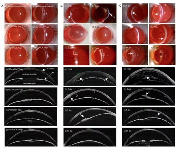
Figure 1 Obvious corneal uveitis appeared in rats after infection with MCMV, manifested as corneal edema, KP and anterior chamber inflammation in varying degrees

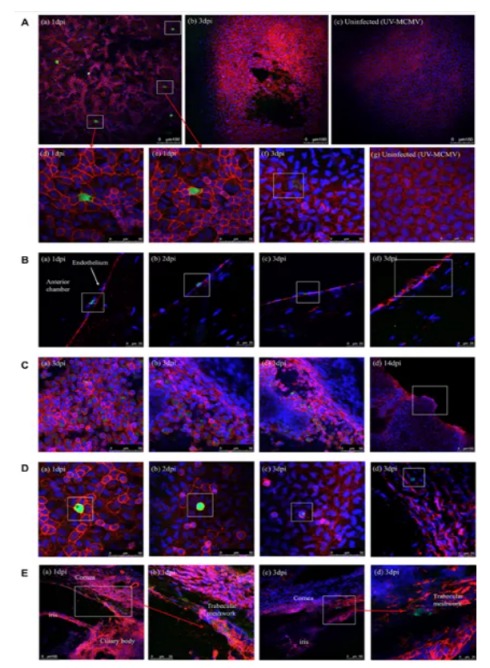
Figure 2 Under a laser confocal microscope, it can be seen that the MCMV-infected anterior segment tissue cells express green fluorescence
Paper link:https://iovs.arvojournals.org/article.aspx?articleid=2778021

Professor Hong Jing
Hong Jing is a second-level professor, chief physician, and doctoral supervisor of Peking University. He is currently the director of the Eye Center, the director of the Department of Corneal and Ocular Surface Diseases, and the director of the eye bank at Peking University Third Hospital. Member of the Ophthalmology Branch of the Chinese Medical Association, Leader of the Ophthalmic Infectology Group of the Ophthalmology Branch of the Chinese Medical Doctor Association, Vice Chairman of the Ophthalmology Branch of the Beijing Medical Association, Vice Chairman of the Ophthalmology Branch of the Chinese Women Physicians Association, Member of the Standing Committee of the Ophthalmology Branch of the Cross-Strait Medical Exchange Committee, Cross-Strait Deputy leader of the ocular surface tear science group of the Ophthalmology Branch of the Medical Communication Committee.
Professor Hong Jing is mainly engaged in the clinical and basic research of corneal and ocular surface diseases. He is the first to carry out corneal endothelial transplantation in China and has been widely promoted in China. In 2014 and 2017, he was selected as one of the “Top Ten Researches in the Field of Corneal Diseases in China in the Past Five Years”. progress". He has long been engaged in basic and clinical research on intraocular viral infections, especially in the diagnosis and treatment of viral diseases in the anterior segment of the eye. He has published a series of articles on the imaging examination, precise diagnosis and treatment of viral corneal lesions, and promoted the virus in my country. The development of sexual corneal disease. Undertook more than 20 key projects of the Ministry of Science and Technology, National Natural Science Foundation of China and other provincial and ministerial projects; wrote 6 professional books "corneal endothelial disease" and published more than 100 academic papers at home and abroad, which were published in "Ophthalmology", "Ocular surface", "AJO", "BJO", "IOVS" and other international authoritative magazines. In 2018, he won the second prize of Scientific and Technological Progress Award for Outstanding Scientific Research Achievements of Higher Education Institutions, and won the Wuzhou Women's Scientific and Technological Progress Award in 2021.